Our website uses cookies to bring you an easy and personalised browsing experience.
By continuing, you consent to our cookie policy on this device. For more info, please see our Privacy Statement.

Accept Cookies
We’re building our community - and our website. New features are being added all the time.
Search results for “Biopsy”
0 results
What happens during a typical prostate biopsy?

For this, tiny samples of prostate tissue (typically 10-12, each measuring around 1mm wide by 2cm long) are extracted using a needle, which can then be examined under the microscope to help confirm or rule out a cancer diagnosis.

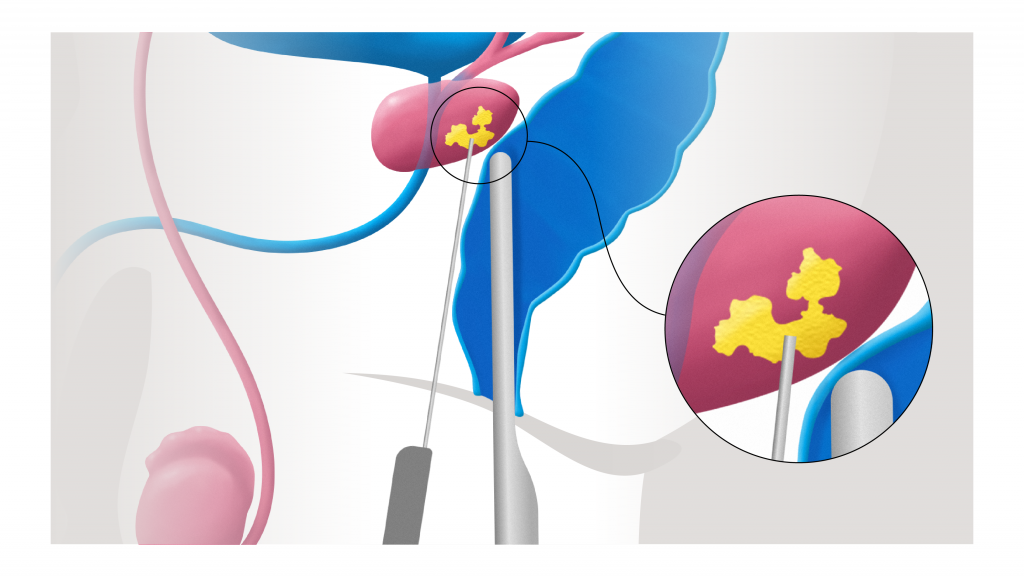
There are different ways of performing a prostate biopsy. The transperineal biopsy, where the needle passes through the perineum, the area of skin between your anus and testicles, is increasingly the most popular option.

For this, you lie on your back and the perineum is numbed with an injection of local anaesthetic. Once that is numbed, local anaesthetic will be injected into the prostate itself.

The biopsy needle is put in place using ultrasound guidance, which means you need to have an ultrasound probe inserted into your rear end during the procedure.

The whole procedure takes around 20 minutes, depending on how many samples are taken.

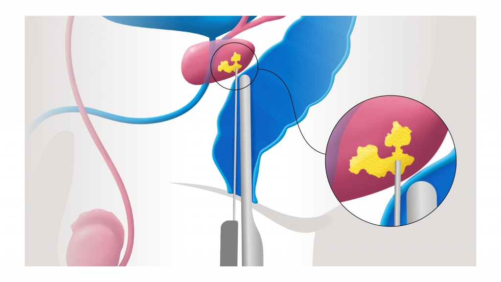
The other option is a transrectal biopsy, the only differences being that you lie on your side and the needle passes through the wall of the back passage, not the perineum. This is now rarely used in the UK and is becoming increasingly less popular in the US, as it has a slightly higher infection risk than a transperineal biopsy.

You will, however, be given antibiotics to take in the days before and after either form of biopsy to reduce the risk of infection.

Are there alternative ways of performing a prostate biopsy?

Around 80 per cent of prostate biopsies are done under local anaesthetic, but in some cases – for example if you found other examinations painful or need a large number of biopsies taken – you may also be offered sedation or a general anaesthetic.

If a previous biopsy has not found any sign of cancer, despite there being a high degree of suspicion that cancer is present, or if your previous biopsy was inconclusive, then you may be offered a transperineal template biopsy, which involves taking more samples than are normally taken during other prostate biopsies. This is done under a general anaesthetic and involves using a grid (or template), which is pressed between the anus and scrotum. The idea is that a biopsy needle can then be used to systematically take a sample from each grid box so that every area of the prostate is sampled. In total, 30-50 samples may be taken.

Does a prostate biopsy hurt?

A biopsy should not be painful, as those not done under general anesthetic are done under local anaesthetic. It will hurt briefly as the local anaesthetic is injected, but this takes only a matter of seconds.

When each sample is taken, you will hear a clicking sound and may feel a flicking sensation.

No one is going to look forward to a biopsy, but remember it will be over in 10-20 minutes and it is the best and most accurate way to detect prostate cancer.

How long will I have to wait for my prostate biopsy results?

This varies according to your hospital or clinic, but they should be available within about a week.

Who will do my biopsy?

This is almost always the job of a urologist. In unusual circumstances, experienced specialist nurses may also perform them.

Will I need to stay in hospital after my biopsy?

No, even if your biopsy is done under general anaesthetic, it is a day case procedure and you should normally be able to go home afterwards.

Are there any risks involved with having a biopsy?

The main risk is infection, which is why you will usually be given antibiotics to take in the days before and afterwards. The risk is, however, far lower with the transperineal biopsy (under 1 per cent) than with other forms of biopsy.

The majority of infections begin within a week of the biopsy. Signs that you have a possible infection include having cloudy or smelly urine, pain in your abdomen or a fever – if you have any of these symptoms in the days following your biopsy, immediately contact your urologist or seek other urgent medical help. Some infections may require treatment in hospital with intravenous antibiotics.

The swelling caused by the procedure may cause some problems urinating in the days afterwards – this is most common after a template biopsy, as more samples are taken than during other biopsies. You will normally be asked to urinate before you go home after your biopsy. If you are later unable to pass urine at home, contact your doctor – you may need a temporary catheter.

You may also notice some blood in your urine (or stools) for a few days afterwards. This is normal – but if it persists beyond a few days or becomes heavier, consult your doctor.

It is also common to have blood in your semen for up to 12 weeks afterwards – which may appear red or rust-coloured. This is not a cause for concern, nor is it dangerous for your partner.

Some men experience temporary erection problems after a biopsy, due to swelling around the prostate affecting the nerves involved in having an erection. However, this only happens in a minority of cases and the issue will normally resolve itself within weeks as the swelling calms down.

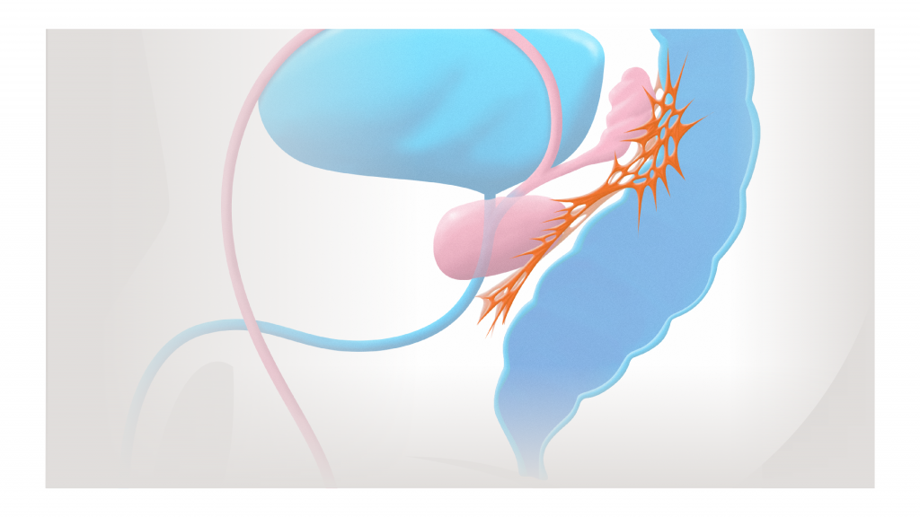
There are two nerves that control erections, with one nerve running down each side of the prostate in the groove between the prostate and the rectum.
How effective is a biopsy at identifying the presence of cancer?

While a biopsy is the most effective way to determine if you have prostate cancer, there is a chance that the presence of cancer may be missed, as the needle samples only the prostate tissue and not any cancer that maybe present.

This is less likely with a template biopsy, which involves taking more samples than other types of biopsy.

A biopsy is the best and most reliable way to identify cancer that requires treating. A biopsy can also identify a cancer that is slow-growing and not aggressive and unlikely to cause any symptoms.

Will I need scans after a biopsy?

If your biopsy has found signs of cancer, a scan may be suggested to see if it has spread beyond the prostate. The type of scan suggested varies but may include a CT scan, an isotope bone scan or a PSMA PET CT scan.

Do I really need to have my cancer genetically tested?

Tests that determine the genetic profile of the cancer can help doctors understand how your cancer is likely to behave and how quickly it might grow or spread.

These tests are used at different stages of the process. Exosome Dx or Select MDx can pick up genetic material from prostate cancer cells in a urine sample before a prostate biopsy. Oncotype MDx tests tissue taken during the biopsy. Another genomic test, Decipher, can be done on either prostate biopsy tissue or prostate tissue removed after surgery.

Genomic tests have limited availability in the UK (where they are available privately) and the US (where they can be expensive). They can, however, give additional useful information to help determine the best form of treatment for you. The best option is to discuss with your urologist whether genomic tests are necessary and which of them is most suitable to your situation.

What difference will scans make to my treatment?

Scans help determine if your cancer is low, intermediate or high-risk – which helps your medical team plan and recommend the appropriate treatment options.

What is a CT scan?

A computerised tomography scan or CT scan helps build images of the internal structures of the body – not just the bones as an X-ray would, but also the blood vessels and soft tissues – and can detect if your cancer has spread elsewhere in the body.

To have a CT scan, you lie on your back and pass through a thin, ring-shaped machine. The scans should be completed in 10-20 minutes. It won’t hurt and, unlike an MRI, does not generate a lot of noise; nor are you enclosed, so it is less likely to make you feel claustrophobic.

What is a PSMA PET CT scan?

This is the most sensitive type of scan for detecting prostate cancer cells, wherever they are in the body. At the moment it is mainly used for those whose cancer has returned after TREATMENT, but the potential for its use is developing all the time.

To have the scan you first have a dye injected which contains radioactive makers which attach to PSMA, a protein found on the surface of prostate cancer cells and then show up on the scan.

PSMA PET CT scans are increasingly used in the UK, but have only recently been approved by the FDA in the USA and so there is limited availability.

We suggest you speak to your urologist about whether you are eligible, and where the scan is available.

The scan takes 20-40 minutes to complete.

What is an isotope bone scan?

An isotope bone scan is performed to check if the cancer has spread to the bones.

For this, a small amount of radioactive dye is injected into your veins, which will gather at areas of abnormality, where bone is breaking down.

The scanner is then passed over your body and will detect these ‘hot spots’. It can take up to an hour to complete.

Who can I discuss my scan results with?

Your urologist or GP will explain and help you to understand the results of your scans.

What happens if I have an abnormal PSA result?

Normally, your doctor will look at your individual circumstances to decide if further tests are necessary. They make this decision taking into account, for example, your age, symptoms and whether there is a family history of prostate cancer. They may decide that the best course of action is to have your doctor recheck your PSA again in six months or a year – only a small proportion of those men who have a raised PSA will have prostate cancer. Or they may decide to send you for an MRI or a biopsy.

What are PSA velocity and density?

The terms may sound complex, but what they represent is straightforward. If you have a large prostate gland – simply through age or swelling, for example – you will most probably have higher levels of PSA anyway. The PSA density calculation accounts for that by measuring the volume of the prostate with ultrasound and dividing that by the PSA level. A high PSA in a small prostate suggests a higher chance of finding cancer at prostate biopsy.

PSA velocity is the rate at which the PSA level climbs. This is similar to PSA doubling time – except it measures the rate of the rise, not the time that it takes to double. The higher the rate, the higher the chance of finding cancer during a biopsy, or the higher the likelihood the cancer has returned after treatment.

Why do I need an MRI?

This is offered if your PSA and other previous assessments suggest that it warrants it. It helps give your doctor a better idea of what is going on in the prostate and helps them decide if you need a biopsy of the prostate.

Unlike a biopsy an MRI is noninvasive, although the machine can be noisy and some men may find lying in the scanner uncomfortable if they don’t like being in confined spaces. However, it should take no more than 30-40 minutes.

What is a PI-RADS score?

This is a score, graded from 1-5, which represents how likely it is that any lesions spotted on your MRI are in fact cancer in the prostate. A score of 1 means it is very unlikely that you have prostate cancer that needs treatment. A score of 5, however, suggests a high risk (70-80 per cent) that you have prostate cancer that needs treatment. A score of 4 means it is likely you have cancer that needs treatment, whereas 3 is sometimes called ‘borderline’ (approx. 20% chance of finding cancer), meaning it is impossible to tell from the result if you have prostate cancer that needs treatment or not. A biopsy will usually be recommended if you have a score of 4 or 5, and sometimes 3, depending on personal circumstance and risk factors.

What is the ‘stage’ of the cancer?

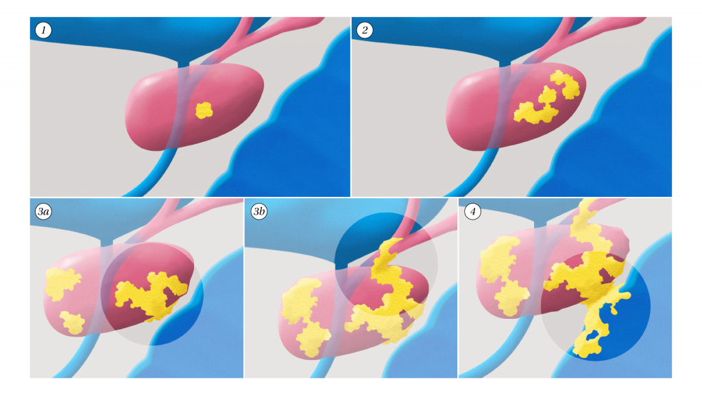
This refers to the size of the cancer and how far it has spread. The stage will normally be represented as a letter T (for tumour), followed by a number from 1 to 4.

T1 means it is so small it may not be seen on a scan.

T2 means it is still enclosed within the prostate.

T3a means it has just broken beyond the perimeter of the prostate, and T3b means it is invading the sperm tubes. Both are referred to as being ‘locally advanced’.

T4 means it has already spread elsewhere surrounding structures such as the rectum OR pelvic wall.

There may also be an ‘N’ number, which reflects whether the cancer has spread to the lymph nodes in the pelvis. – Zero (N0) means no nearby lymph nodes are affected; 1 (N1) signifies that the cancer has spread to one or more nodes.

The “M” number refers to the presence of metastasis – M0 means no metastasis present and M1 means metastasis present.

Fig 1: T1 refers to prostate cancer that is localised to the prostate, in a small proportion of one half of the gland. The original classification of T1 was complicated and split into 1a, 1b and 1c. 1a and 1b were diagnosed after a TURP operation, and 1c was diagnosed after screening biopsy but this has been changed after the introduction of MRI.
Fig 2: T2 means it is still enclosed within the prostate, but involving either both halves of the prostate or more than 50% of one half.
Fig 3a: T3a means it has just broken through the capsule of the prostate.
Fig 3b: T3b means it is invading the sperm tubes.
Fig 4: T4 means invading surrounding structures beyond the sperm tubes such as the pelvic side wall or the rectum. T3 and T4 are referred to as locally advanced.
What are the Gleason grade and score?

The Gleason grade measures the potential aggression of the cancer, which is determined after a pathologist has looked at samples of your prostate cancer under the microscope.

The grade goes from 1-5, with 1 indicating cancer that is going to grow very slowly, if at all, and 5 being the highest, meaning the cancer is likely to grow quickly.

To give the most accurate picture, two scores are given – the first representing the most common type of cells found in the biopsy, and the second number being the highest grade of the remaining cells. The two are added together to give a total score. So, if most of the cells were a Gleason grade 3 and the rest were mainly a 4, this would give 3 + 4 (and a total score of 7).

Generally, a total score of 6 or less means the cancer is likely to grow slowly if at all, 7 indicates an intermediate risk cancer with a moderate risk of growth, and above 7 means it is a high risk cancer likely to grow quickly.

Your Urologist may also refer to another grading system known as the ‘Gleason Grade Groups’ (GGG), which also runs on a range from 1 to 5. GGG1 is the equivalent of total Gleason score of 6; GGG2 equates to Gleason 3 +4; GGG3 to Gleason 4+3; GGG4 to Gleason 4+4; and GGG5 to a total Gleason score of 9 or 10.

I have been offered active surveillance. What does that mean?

You are usually offered this option if the tests conducted suggest that you have low-risk, slow-growing cancer that is confined to the prostate.

It means that rather than having any active treatment, such as radiotherapy or surgery, your cancer will be monitored to see if it progresses.

You will typically have a PSA blood test every three months, an annual MRI and a repeat biopsy about 2 years after your initial biopsy. The MRI may be repeated more frequently if your PSA climbs quickly.

Is active surveillance safe?

As we understand more and more about prostate cancer, it is clear that low-risk disease (defined by a Gleason score of 3+3 on your biopsy) is very different to intermediate or higher risk disease. Defining it as cancer is arguably inaccurate, as cancer cells normally grow and multiply uncontrollably – whereas low risk prostate cancer cells multiply very slowly.

As the risk posed by such cancer is so low, the internationally approved recommendation is that active surveillance is the right treatment. The low risk of this type of cancer does not warrant the potential risks that can come with surgery or radiotherapy.

I don’t want to live with untreated cancer. Are there other options?

It really depends on your biopsy and staging results. If your Gleason score was 3 + 3 and it is clear from tests that you have low-risk disease, it is highly unlikely that you will be offered surgery or radiotherapy, even if you request it.

The recommendations for active surveillance for low-risk disease are determined by international convention and it is also highly unlikely that a reputable doctor will offer treatment that goes beyond those guidelines.

How often do I need a biopsy with active surveillance?

This varies from centre to centre, but you will normally have a repeat biopsy one or two years after your first. Further biopsies after this may be required if there are any changes in your physical examination, PSA, or MRI results.

Which nomogram is best?

No one nomogram is better than the other.

Some may suit some patients better, because they are directed at different stages of diagnosis or treatment – for example, after biopsy when considering which treatment to have, or when considering further radiotherapy.

Just ensure the one you use is supported by a reputable institution such as a university or hospital.

search-icon
There were not matches for your search. Try reviewing your search or filter criteria.
Other search results include
Our site is under development right now and our community is growing.
Please sign up for our newsletter and be the first to know about the latest developments and our progress.

    NowWhat?
    Thank you for subscribing you will now receive a welcome email. Please check your inbox and if you don’t see our message please check your spam or junk folders.
    Contact us

      Thank you!
      We will contact you as soon as possible!
      NowWhat?
      Thank you for subscribing you will now receive a welcome email. Please check your inbox and if you don’t see our message please check your spam or junk folders.
      Become a mentor
      Please fill in the form below and one of our team will be in touch

        Thank you
        We thank you sincerely for wanting to help
        Set up fundraising
        Please fill in the form below and one of the team at Race Nation will be in touch to guide you through the process

          Thank you
          The Race Nation team will be in touch soon
          Create an event
          Please fill in the form below and one of the team at Race Nation will be in touch to guide you through the process

            Thank you
            The Race Nation team will be in touch soon
            Breast Cancer Community
            We are developing a sister site for breast cancer which is expected to be launched in 2023.
            NowWhat?
            Dr. NowWhat
            Benefit from Dr. NowWhat’s 30 years of clinical experience treating patients
            Hello, I’m Dr. NowWhat, I’m here to help!
            What are you wondering about?
            Register
            Please register as a patient, former patient, or a patient supporter
            Already have an account? Log in
            Please read our Terms of Service before you proceed with the registration.
            Your account successfully created!
            Check your email to continue registration!
            Welcome back!
            Don’t have an account yet? Register
            Forgot password?
            Your account successfully created!
            Check your email to continue registration!
            Account is deactivated
            Your account has been deactivated and is queued for deletion Do you want to restore it?
            Success
            Your account was successfully restored!
            Forgot password?
            Send a link to your email to reset password.
            Check email
            You received a link in your email to reset password.
            Your email successfully confirmed!