
A revolutionary new scan that makes tumour cells ‘glow’ could make it much easier for doctors to detect prostate cancer.
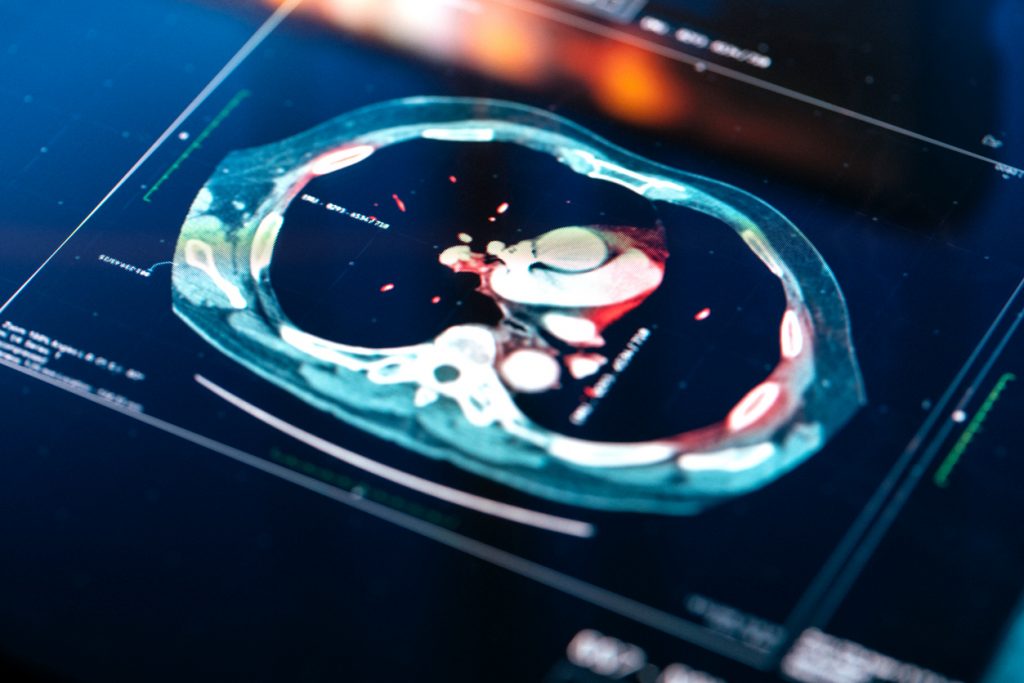
The high-tech scan is a new form of Magnetic Resonance Imaging (MRI) that makes malignant cells light up on the computer screen, while healthy tissue does not.
This makes it easier for medics to identify cancerous tissue and track its growth over time.
Scientists who developed the technology at the University of Waterloo in Ontario, Canada, hope it will improve detection rates and help doctors spot prostate cancer at an earlier stage – when it is more treatable and there is a better chance of defeating it.
MRI scans are often used as part of the diagnostic process in prostate cancer.
They use a magnetic field, combined with radiofrequency pulses, to create detailed pictures of what’s going on inside of the body.
This helps doctors to evaluate the extent of any prostate tumour present and to work out over time whether it has spread.
The advantage is that it can help men avoid having an unnecessary biopsy – if the scan is clear, there is usually no need to have prostate tissue surgically removed for testing.
And if a biopsy is needed, the MRI scan allows doctors to target the part of the prostate where cancer seems to be present.
But sometimes an MRI can wrongly diagnose prostate cancer when the real cause may be inflammation due to something else such as prostatitis.
Making tumour cells light up could improve the accuracy of the scan.
The technique developed by the Waterloo team is based on the way water molecules move around in cancerous tissue.
Cancer cells tend to be more misshapen and irregularly packed together than non-cancerous ones which disrupts the normal flow of water molecules inside the tissue.
The new test capitalises on this by analysing the way irregular movement of water disrupts MRI signals.
It converts those areas where disruption is detected into ‘glowing’ hot spots on the scan.
The technology was tested on 200 men with prostate cancer and was found to be significantly more accurate at spotting cancerous tissue than standard MRI, according to findings published in the journal Scientific Reports.
Research leader Alexander Wong, professor of systems design engineering at the University of Waterloo, said: ‘This new technology has promising potential to improve cancer screening, prognosis and treatment planning.
‘We also have very promising results for breast cancer.
‘This could be a game-changer.’